Open Access
Figure 5.
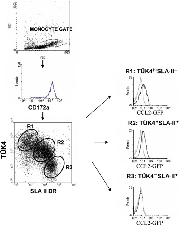
Differential binding of CCL2-GFP to monocyte subsets. Porcine PBMC were incubated with CCL2-GFP (CHO cell supernatant) followed by Alexa 488-conjugated rabbit anti-GFP IgG, and then double stained for Tük4 and SLA II. The histograms are gated on the appropriate monocyte subpopulation regions as defined in the dot-plots. Grey histograms show the background fluorescence of monocytes incubated with supernatants from CHO cells transfected with the InvCCL2-GFP construct. 25 000 cells were acquired. (A color version of this figure is available at www.vetres.org.)