Figure 2.
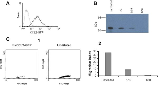
Expression of recombinant porcine CCL2. (A) CHO cell line stably expressing the porcine CCL2 fused to GFP. The expression of GFP fusion protein was directly analysed by flow cytometry. Non transfected CHO cells were used as negative control (grey histogram). 5 000 cells were acquired. (B) Western blot of CCL2-GFP produced by transfected CHO cells. Different dilutions of supernatant were resolved by 15% SDS-PAGE under reducing conditions and revealed with biotinylated anti-GFP and streptavidin-HRP. Numbers on the left indicate the position of MW markers. (C) Chemotactic activity of CCL2-GFP on porcine blood monocytes. Chemotaxis was assessed with the Transwell cell migration system and subsequent flow cytometry counting of migrated cells by a 45 s acquisition. (1) FSC versus SSC dot plot of migrated cells in response to supernatants from CHO cells expressing CCL2-GFP or the inverted sequence of pCCL2 fused to GFP (InvCCL2-GFP, negative control). (2) Results expressed as migration index, calculated as the ratio of the number of cells migrating to the chemokine and the number of cells in the negative control. Results from one representative experiment out of three performed are shown. (A color version of this figure is available at www.vetres.org.)