Figure 1.
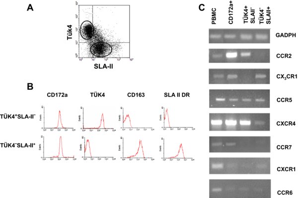
Chemokine receptor mRNA expression in monocyte subsets. (A) Dot plot of Tük4 versus SLA-II expression in monocytes obtained from PBMC (20 000 cells were acquired). Three subsets can be identified: Tük4+SLA-II−, Tük4+SLA-II+, and Tük4−SLA-II+. (B) The Tük4+SLA-II− and Tük4−SLA-II+ monocyte subsets were magnetically isolated as described in Materials and methods section, and the expression of the indicated markers was analysed by flow cytometry (5 000 cells acquired). (C) Total RNA isolated from PBMC, the whole monocyte population (CD172a+) or the Tük4+SLA-II− and Tük4−SLA-II+ monocyte subsets shown in (B) were reverse transcribed, and the cDNA amplified by PCR with specific primers for GAPDH or different chemokine receptors. PCR products were analysed by 1.5% agarose gel electrophoresis. Data are representative from three independent experiments using different donors. (A color version of this figure is available at www.vetres.org.)