Figure 4.
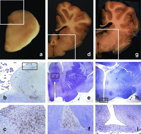
Accumulation of PrPd in CVO and related brain structures. a, d, g: Gross sections showing location of the AP at the level of the obex (a), of the SFO at the level of the anterior hypothalamus (d), and of the ME at the level of the infundibular hypothalamus (g). b, e, h: Low power micrographs corresponding to the framed areas of a, d and g, respectively. PrPd is seen to accumulate in the framed areas that correspond to the AP (b), SFO (e) and ME (h); IHC with Bar224 PrP antibody and haematoxylin counterstaining (b, ×2; e, ×1; h, ×1). c, f, i: Detail of PrPd accumulating in the same CVO as in b, e and h, respectively; IHC with Bar224 PrP antibody and haematoxylin counterstaining (c, ×10; f, ×4; i, ×4). (For a color version of this figure, please consult www.vetres.org.)