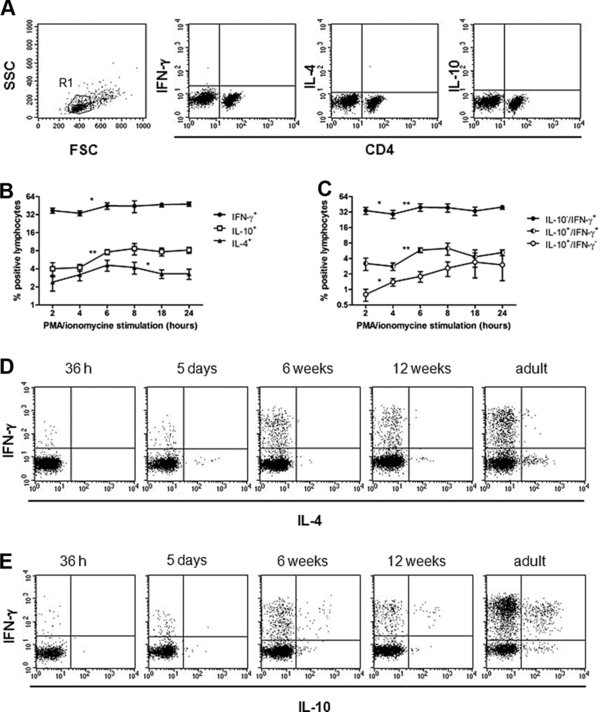
Figure 1.

Flow cytometric analysis of IFN-γ, IL-4 and IL-10 producing PBMC after stimulation with PMA and ionomycin. PBMC were incubated in the presence of the secretion blocker Brefeldin A. Then, they were fixed and intracellular cytokine staining was performed. (A) Non-stimulated PBMC from a 6 week old foal after 4 h of incubation in medium with Brefeldin A. The left plot shows the gating (R1) on peripheral blood lymphocytes that was used for analysis of the data. The remaining three plots show a two-color staining of non-stimulated lymphocytes using anti-CD4 and different anti-cytokine antibodies. Typically, cytokines were not detected in equine peripheral blood lymphocytes in the absence of stimulation. Isotype controls for cytokine staining generally resulted in less than 0.05% of detectable cells. (B and C): PBMC from 4 adult horses were stimulated with PMA and ionomycin for up to 24 h and cytokine expression was measured at various time points. (B) Total percentages of IFN-γ, IL-10 and IL-4 producing cells in the lymphocyte population, (C) Percentages of IFN-γ+/IL-10+, IFN-γ+/IL-10− and IFN-γ−/IL-10+ cells during 24 h of stimulation. The data in B and C represent means and standard deviations. Differences in cytokine expression from one to the next time point were compared by Student’s t-tests. ** p = 0.001 to < 0.01; * p = 0.01 to 0.05; (D) PMA and ionomycin stimulated cells from foals of different ages and adult horses. Double staining of IFN-γ and IL-4 or (E) IFN-γ and IL-10. The dot plots show one representative image from a foal or horse of the respective age group.


