Figure 3.
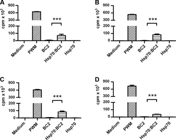
CD4+ T cell proliferation induced by MHC-matched monocytes to Hsp70 complexes. (A–D) Monocytes isolated from naïve A31 homozygous cattle were co-incubated with CD4+ T cells from FMD17 (A31/A14) and either FMDV 25-mer peptide “BC2” (5 ng/mL), Hsp70 (1 μg/mL) or BC2 and Hsp70, at the same final concentrations, pre-incubated to form a complex. Responses to Pokeweed mitogen (PWM) are also indicated. After 5 days, wells were pulsed with 37 kBq [3H] thymidine and incubated for a further 16 h before harvesting. Incorporated radioactivity was determined by liquid scintillation counting and expressed as counts per minute (cpm). Data are presented as the cpm × 103/min mean ± S.D. of triplicate cultures. One representative data set of three is shown for each of the four animals. Significant differences in proliferation between BC2 and Hsp70:BC2 stimulated cells are indicated (*** p < 0.001).