Open Access
Figure 1.
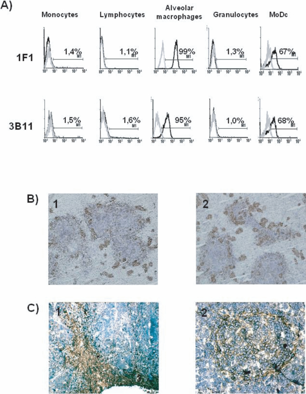
(A) MAb 1F1 and 3B11 recognize an antigen expressed in alveolar macrophages and MoDC. Cells were labeled with 1F1 or 3B11 mAb (black histograms) or with irrelevant isotype-matched mAb (grey histograms). Results are representative of five independent experiments. (B) In the spleen, staining with 1F1 (1) and 3B11 (2) mAb appears associated with the walls of the ellipsoidal vessels that surround the islands of white pulp. (C) In lymph nodes, the antigen was detected in the subcapsular sinus and medullary cords (1) and in the periphery of follicles and some follicular cells (2). (A color version of this figure is available at www.vetres.org.)