Free Access
Figure 5.
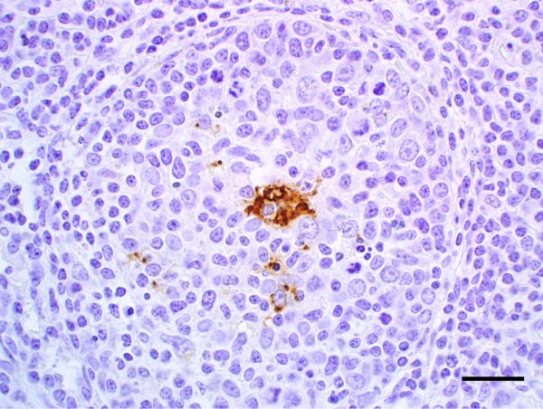
Lymph node, pig, inoculated with porcine circovirus type 2 (PCV2) 140 days previously. Immunohistochemical staining using a polyclonal antiserum against PCV2 revealing scant brown staining in the cytoplasm of macrophages-like cells in the center of lymphoid follicles. Streptavidin-biotin-peroxidase complex method counterstained with hematoxylin. Bar = 40 μm. (A color version of this figure is available at: www.vetres.org.)