Free Access
Figure 4.
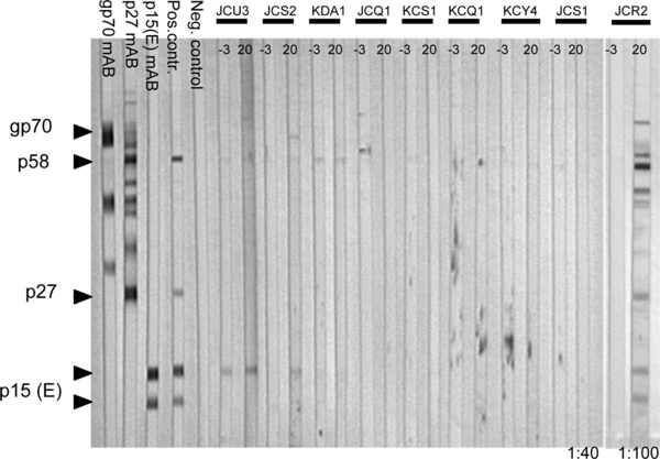
Western blot analysis using plasma samples from week −3 (prior to challenge) and week 20 p.i. (cats group 100K and two of group 10K). The first 3 strips to the left were incubated with monoclonal antibodies against gp70, p27 and p15(E), respectively, to characterize the respective viral proteins. Serum obtained from a pool of immune cats used as a positive control. Cat JCU3 and cat JCS2 (group 10K) show a weak reaction with the upper band of the p15(E) protein. JCR2 shows antibodies to all FeLV proteins. In all other cats, responses to any FeLV proteins could not be detected. (▶) bands with expected length. All samples were tested under the same assay conditions.