Free Access
Figure 4.
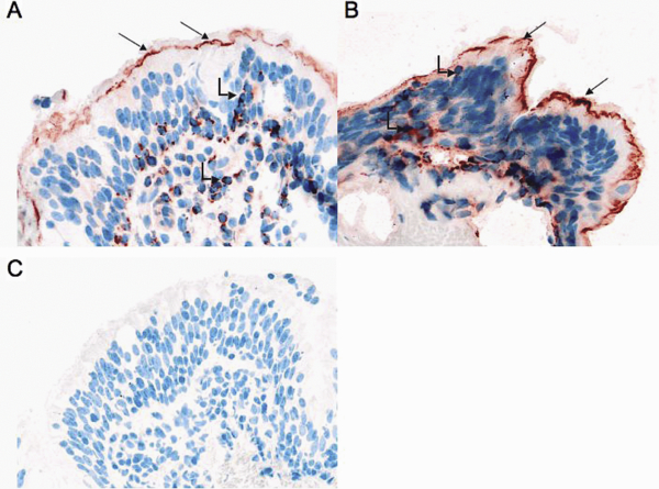
PTX3 expression in tissue samples. PTX3 was constitutively expressed in bronchial epithelial cells (straight arrows) and was localized at the apex just behind ciliated processes. It was also detected in infiltrative inflammatory cells (angled arrows). (A) Bronchial section from a healthy horse. (B) Bronchial section from a horse suffering from R.A.O. with over-expression of PTX3 in bronchial epithelial cells and an increase of the number of infiltrative inflammatory cells expressing PTX3. (C) Control experiment carried out using an IgG2β isotype showed no staining of PTX3 (the same results were obtained by omitting the primary antibody). Magnification ×200. (A color version of this figure is available online at www.vetres.org.)