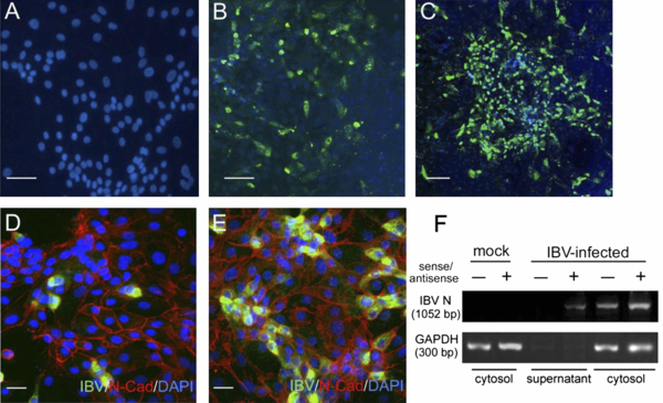
Figure 3.

Infection of ATE cells with IBV. A total of 5 × 10⁴ ATE cells were either non-infected (A) or infected with 50 μL of 2575/98 strain (EID50 = 105/mL) for 1 h at 37 °C. At 24 h.p.i. (B, D) and 72 h.p.i. (C, E), the expression of IBV proteins was detected by chicken anti-serum against IBV (200-fold diluted). Infected cells were further characterized by the staining for E-cadherin (red) (D, E). (F) Total RNA was isolated from the supernatants and cell lysates of infected and uninfected ATE cells at 24 h.p.i. The cDNA derived from viral sense (+) and antisense RNA (−) were detected by primers targeting to the nucleocapsid (N) gene. The house-keeping gene GAPDH was used as an internal control. Scar bar in panel (A) to (C), 25 μm; in panel D and E, 10 μm. (A color version of this figure is available at www.vetres.org.)


