Open Access
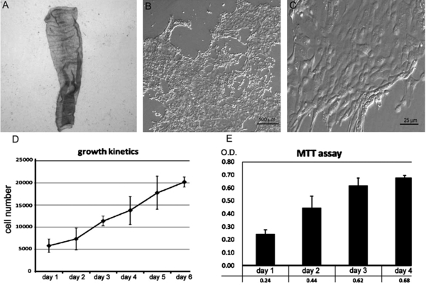
Figure 1.

Morphology and growth curve of primary avian tracheal epithelial (ATE) cells. (A) An isolated intact membrane sheet of the tracheal epithelium from a one-day-old chick. After digestion with collagenase I, the dissociated ATE cells were plated on 2% matrigel-coated 24-well plates. The morphology of the ATE cells are shown in panel (B) and (C). The cell growth was further analyzed by trypan blue exclusion assay (D) and MTT activity (E). Y-axis (D) states the cell number per well of 24-well plates.


