Free Access
Figure 1.
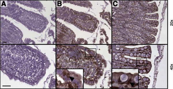
Immunohistochemical detection of CD3+ cells in the small and the large canine intestine. Paraffin sections of canine small and large intestine were stained with control rabbit IgG (A) or anti-CD3 antibody (B, C). Lower (20×) and higher magnifications (40×) of representative stainings of small (A, B) and large intestine (C) are shown. Inserts show enlargements of the epithelial layer with IEL (arrow). Bars indicate 50 µm.