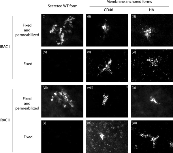
Figure 3.

Immunodetection of IRAC expressed by BoHV-4 recombinant strains. MDBK cells were infected at a MOI of 0.01 PFU/cell with BoHV-4 V. test IRAC I (i) and (iv), BoHV-4 V. test IRAC II (vii) and (x), BoHV-4 V. test BAC G IRAC I CD46 (ii) and (v), V. test BAC G IRAC I HA (iii) and (vi), V. test BAC G IRAC II CD46 (viii) and (xi) and V. test BAC G IRAC II HA (ix) and (xii) excised strains. After 48 h, cells were fixed and permeabilized (panels (i–iii) and (vii–ix)) or only fixed (panels (iv–vi) and (x–xii)), then treated as described in the methods for indirect immunofluorescent labeling. mAbs anti-IRAC I or II were used as primary antibodies and were revealed by Alexa488-GAM secondary antibodies. The side of each panel corresponds to 500 μm of the specimen.


