Free Access
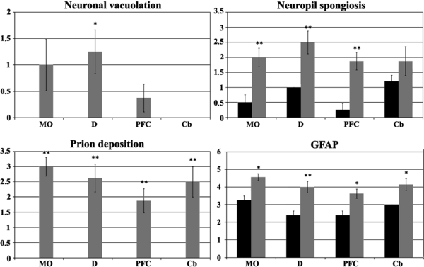
Figure 2.

Scoring of histopathological lesions in each analysed area. Scores for neuronal vacuolation, neuropil spongiosis, prion deposition and reactive gliosis (GFAP) are shown as means ± standard error. Black bars: control sheep, grey bars: scrapie-infected sheep. Areas: medulla oblongata (MO), diencephalon (D), prefrontal cortex (PFC), and cerebellum (Cb). Student t-test * P < 0.05, ** P < 0.01.


