Free Access
Figure 2.
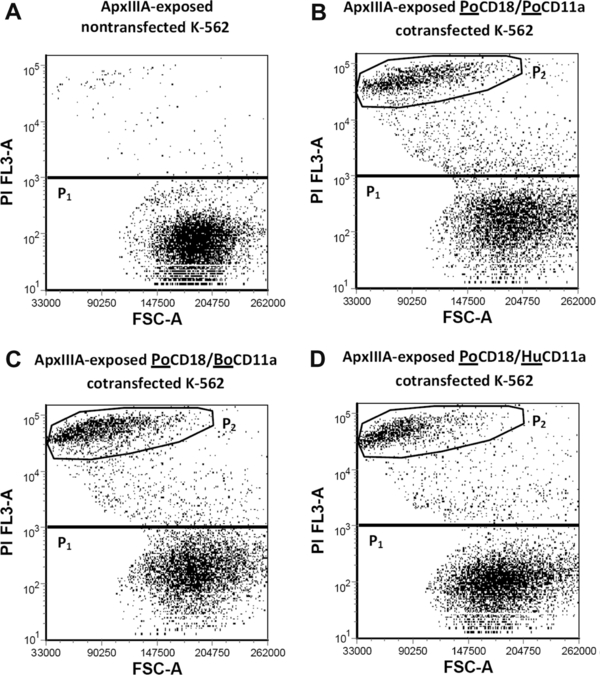
K-562 cells accumulate PI upon ApxIIIA exposition when expressing porcine CD18-containing LFA-1s. PI versus forward scatter (FSC) dot plot from flow cytometric analysis of ApxIIIA-exposed control (top, left) and double transfected K-562 cells. Only the P2 populations expressed the intended LFA-1.