Figure 2.
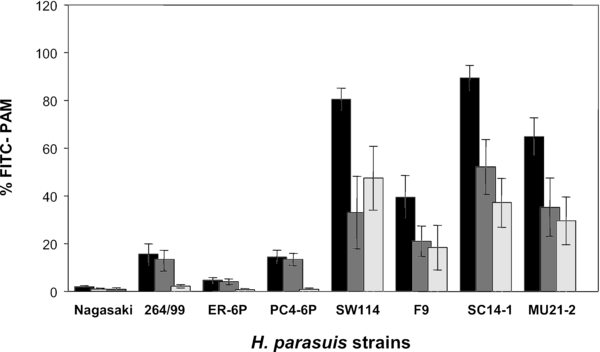
Phagocytosis of H. parasuis strains by PAM. PAM (5 × 10⁵ cells/well) were incubated with different FITC-labelled strains of H. parasuis. After 1 h of incubation, extracellular bacteria were detected with a H. parasuis-specific antiserum coupled with a phycoerythrin-secondary antibody. Macrophages were then analysed by flow cytometry. Results are presented as the percentages of cells associated with fluorescent bacteria (black bars), cells with bacteria inside (only green fluorescence; dark gray bars) and cells with bacteria on the surface (green and red fluorescence; light gray bars). Inoculi of bacteria were: 109 CFU/well of Nagasaki; 2–4 × 10⁸ CFU/well of 264/99, ER-6P and PC4-6P; 7–8 × 10⁷ CFU/well of SW114, F9 and SC14-1. Data are means and standard deviations from four independent experiments.