Free Access
Figure 5.

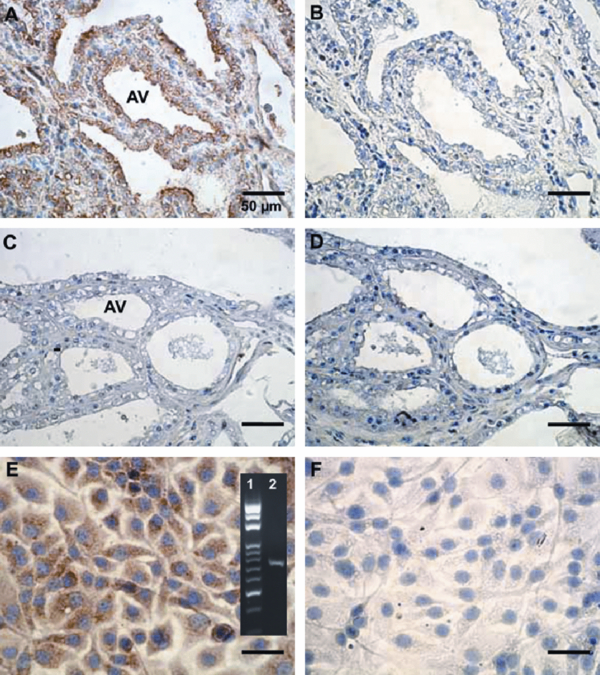
Immunocytochemical localization of Hp in mammary gland and MAC-T cells. Mammary tissues with (A and B) and without mastitis (C and D) were incubated with mouse anti-Hp polyclonal antibodies (A and C), while using normal non-immuned mouse serum as a negative control (B and D). MAC-T cells were incubated with mouse anti-Hp polyclonal antibodies (E), while using normal non-immuned mouse serum as a negative control (F). DAB was used as a chromogenic substrate for Hp staining (in brown) and hematoxylin was employed as a nuclear stain (in blue). Panel in E represents the Hp mRNA expression (Lane 2) and DNA molecular weight markers (Lane 1) using RT-PCR. AV = alveolar.


