Free Access
Figure 1.
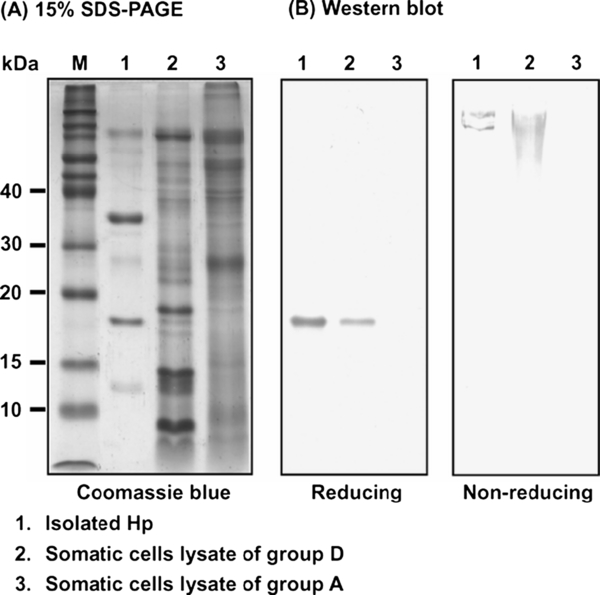
Typical SDS-PAGE pattern and Western blot analyses of milk somatic cell lysates from groups A and D. (A) Coomassie blue staining of somatic cell lysates run on a 15% SDS-PAGE under reducing conditions. (B) Western blot analysis of the isolated Hp and somatic cell lysates under reducing (left panel with 15% SDS-PAGE) and non-reducing conditions (right panel with 4% SDS-PAGE) using a mouse polyclonal antibody prepared against bovine recombinant Hp. Lane M, molecular markers in kDa.